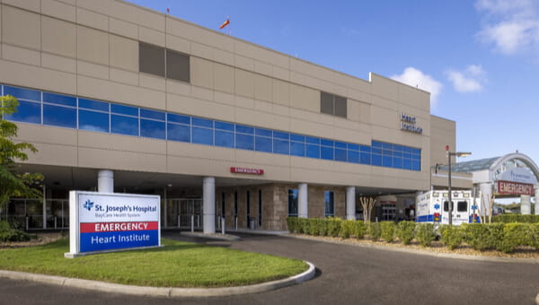
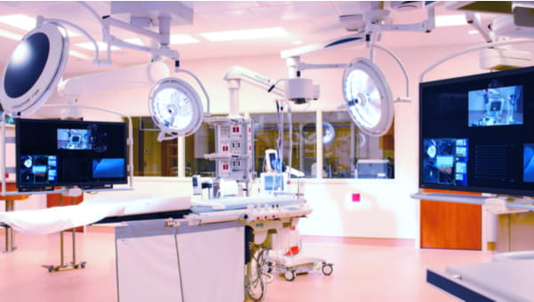
As one of the nation’s Top 50 cardiovascular hospitals as recognized by Premier Inc.’s 50 Top Cardiovascular Hospitals®, St. Joseph’s Hospital has everything your heart desires. Premier Inc. is a trusted source that performs an annual research study that ranks the top hospitals in the country. At St. Joseph’s Hospital, we provide a continuum of high-quality care and services that save lives and improve the well-being of the people in our communities. St. Joseph’s Hospital’s Heart and Vascular Institute offers cardiac and vascular care services in one location to comprehensively treat all aspects of adult and pediatric cardiovascular disease, from diagnosis to treatment to recovery. We have advanced hybrid technology to meet the growing demand for minimally invasive heart and vascular surgery, as well as additional updated patient rooms.
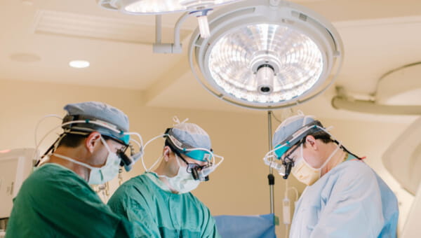
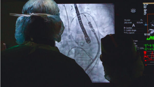
Our Heart and Vascular Institute offers the latest technologies for open-heart and structural heart procedures. Advanced and minimally invasive procedures include complex valve and coronary bypass surgery, TAVR, MitraClip, extracorporeal membrane oxygenation, vascular surgery, targeted hypothermia ablation of advanced atrial fibrillation (AFib) and complex arrhythmia, as well as a complete suite of offerings to manage implantable cardiac devices. Each year, over 500 open-heart surgeries, 600 interventional procedures, 900 diagnostic services and 11,500 diagnostic tests are performed as our nationally renowned heart program continues to grow. But no matter how many procedures we do or awards we receive, what makes us special is how we care for you.